Development of titanium alloys and surface treatments to increase the implants lifetime
DOI:
https://doi.org/10.3989/revmetalm.084Keywords:
Anodization, β titanium alloys, Biocompatibility, Nanotubes, Osseointegration, Surface treatments, Thermal treatmentsAbstract
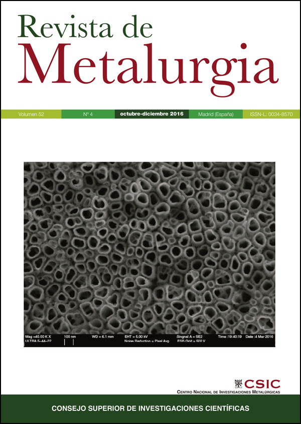
The population aging together with increase of life expectancy forces the development of new prosthesis which may present a higher useful life. The clinical success of implants is based on the osseointegration achievement. Therefore, metal implants must have a mechanical compatibility with the substituted bone, which is achieved through a combination of low elastic modulus, high flexural and fatigue strength. The improvement, in the short and long term, of the osseointegration depends on several factors, where the macroscopic design and dimensional, material and implant surface topography are of great importance. This article is focused on summarizing the advantages that present the titanium and its alloys to be used as biomaterials, and the development that they have suffered in recent decades to improve their biocompatibility. Consequently, the implants evolution has been recapitulated and summarized through three generations. In the recent years the interest on the surface treatments for metallic prostheses has been increased, the main objective is achieve a lasting integration between implant and bone tissue, in the shortest time possible. On this article various surface treatments currently used to modify the surface roughness or to obtain coatings are described it; it is worthy to mention the electrochemical oxidation with post-heat treated to modify the titanium oxide crystalline structure. After the literature review conducted for prepare this article, the ? titanium alloys, with a nanotubes surface of obtained by electrochemical oxidation and a subsequent step of heat treatment to obtain a crystalline structure are the future option to improve long term biocompatibility of titanium prostheses.
Downloads
References
Ahmed, T., Rack, H.J. (1998). Phase transformations during cooling in ?+? titanium alloys. Mat. Sci. Eng. A-Struct. 243 (1-2), 206–211. https://doi.org/10.1016/S0921-5093(97)00802-2
Anselme, K., Bigerelle, M., Noel, B., Dufresne, E., Judas, D., Iost, A., Hardouin, P. (2000). Qualitative and quantitative study of human osteoblast adhesion on materials with various surface roughnesses. J. Biomed. Mater. Res. 49 (2), 155–166. https://doi.org/10.1002/(SICI)1097-4636(200002)49:2<155::AID-JBM2>3.0.CO;2-J
Bai, Y., Park, I.S., Park, H.H., Lee, M.H., Bae, T.S., Duncan, W., Swain, M. (2011). The effect of annealing temperatures on surface properties, hydroxyapatite growth and cell behaviors of TiO2 nanotubes. Surf. Interface Anal. 43 (6), 998–1005. https://doi.org/10.1002/sia.3683
Ban, S., Iwaya, Y., Kono, H., Sato, H. (2006). Surface modification of titanium by etching in concentrated sulfuric acid. Dent. Mater. 22 (12), 1115–1120. https://doi.org/10.1016/j.dental.2005.09.007 PMid:16375960
Bauer, S., Pittrof, A., Tsuchiya, H., Schmuki, P. (2011). Size-effects in TiO2 nanotubes: Diameter dependent anatase/rutile stabilization. Electrochem. Commun. 13 (6), 538–541. https://doi.org/10.1016/j.elecom.2011.03.003
Bayram, C., Demirbilek, M., Yalçin, E., Bozkurt, M., Do?an, M., Denkba?, E.B. (2014). Osteoblast response on co-modified titanium surfaces via anodization and electrospinning. Appl. Surf. Sci. 288, 143–148. https://doi.org/10.1016/j.apsusc.2013.09.168
Berger, S., Hahn, R., Roy, P., Schmuki, P. (2010). Self-organized TiO2 nanotubes: Factors affecting their morphology and properties. Phys. Status Solidi B 247 (10), 2424–2435. https://doi.org/10.1002/pssb.201046373
Berger, S., Albu, S.P., Schmidt-Stein, F., Hildebrand, H., Schmuki, P., Hammond, J.S., Reichlmaier, S. (2011). The origin for tubular growth of TiO2 nanotubes: A fluoride rich layer between tube-walls. Surf. Sci. 605 (19-20), L57–L60. https://doi.org/10.1016/j.susc.2011.06.019
Bjursten, L.M., Rasmusson, L., Oh, S., Smith, G.C., Brammer, K.S., Jin, S. (2010). Titanium dioxide nanotubes enhance bone bonding in vivo. J. Biomed. Mater. Res.- A 92A (3), 1218–1224.
Brammer, K.S., Oh, S., Cobb, C.J., Bjursten, L.M., Heyde, H. Van Der, Jin, S. (2009). Improved bone-forming functionality on diameter-controlled TiO2 nanotube surface. Acta Biomater. 5 (8), 3215–3223. https://doi.org/10.1016/j.actbio.2009.05.008 PMid:19447210
Browne, M., Gregson, P.J. (2000). Effect of mechanical surface pretreatment on metal ion release. Biomaterials 21 (4), 385–392. https://doi.org/10.1016/S0142-9612(99)00200-8
Çali?kan, N., Bayram, C., Erdal, E., Karahalilo?lu, Z., Denkba?, E.B. (2014). Titania nanotubes with adjustable dimensions for drug reservoir sites and enhanced cell adhesion. Mat. Sci. Eng. C 35, 100–105. https://doi.org/10.1016/j.msec.2013.10.033 PMid:24411357
Chlebus, E., Ku?nicka, B., Kurzynowski, T., Dyba?a, B. (2011). Microstructure and mechanical behaviour of Ti-6Al-7Nb alloy produced by selective laser melting. Mater. Charact. 62 (5), 488–495. https://doi.org/10.1016/j.matchar.2011.03.006
Choe, H.C., Kim, W.G., Jeong, Y.H. (2010). Surface characteristics of HA coated Ti-30Ta-xZr and Ti-30Nb-xZr alloys after nanotube formation. Surf. Coat. Tech. 205 (Suppl. 1), S305–S311. https://doi.org/10.1016/j.surfcoat.2010.08.020
Cochran, D.L., Schenk, R.K., Lussi, A., Higginbottom, F.L., Buser, D. (1998). Bone response to unloaded and loaded titanium implants with a sandblasted and acid-etched surface: A histometric study in the canine mandible. J. Biomed. Mater. Res. 40 (1), 1–11. https://doi.org/10.1002/(SICI)1097-4636(199804)40:1<1::AID-JBM1>3.0.CO;2-Q
Cremasco, A., Osório, W.R., Freire, C.M., Garcia, A., Caram, R. (2008). Electrochemical corrosion behavior of a Ti-35Nb alloy for medical prostheses. Electrochim. Acta 53 (14), 4867–4874. https://doi.org/10.1016/j.electacta.2008.02.011
Cremasco, A., Messias, A.D., Esposito, A.R., Duek, E.A.D.R., Caram, R. (2011). Effects of alloying elements on the cytotoxic response of titanium alloys. Mat. Sci. Eng. C 31 (5), 833–839. https://doi.org/10.1016/j.msec.2010.12.013
Das, K., Bose, S., Bandyopadhyay, A. (2007). Surface modifications and cell-materials interactions with anodized Ti. Acta Biomater. 3 (4), 573–585. https://doi.org/10.1016/j.actbio.2006.12.003 PMid:17320494
Das, K., Bose, S., Bandyopadhyay, A. (2009). TiO2 nanotubes on Ti: Influence of nanoscale morphology on bone cell-materials interaction. J. Biomed. Mater. Res.-A 90A (1), 225–237. https://doi.org/10.1002/jbm.a.32088 PMid:18496867
Diniz, M.G., Soares, G.A., Coelho, M.J., Fernandes, M.H. (2002). Surface topography modulates the osteogenesis in human bone marrow cell cultures grown on titanium samples prepared by a combination of mechanical and acid treatments. J. Mater. Sci. - Mater. M. 13 (4), 421–432. https://doi.org/10.1023/A:1014357122284
Duraccio, D., Mussano, F., Faga, M.G. (2015). Biomaterials for dental implants: current and future trends. J. Mater. Sci. 50 (14), 4779–4812. https://doi.org/10.1007/s10853-015-9056-3
Eisenbarth, E., Velten, D., Müller, M., Thull, R., Breme, J. (2004). Biocompatibility of ?-stabilizing elements of titanium alloys. Biomaterials 25 (26), 5705–5713. https://doi.org/10.1016/j.biomaterials.2004.01.021 PMid:15147816
Ferreira, C.P., Gonçalves, M.C., Caram, R., Bertazzoli, R., Rodrigues, C.A. (2013). Effects of substrate microstructure on the formation of oriented oxide nanotube arrays on Ti and Ti alloys. Appl. Surf. Sci. 285 (Part B), 226–234. https://doi.org/10.1016/j.apsusc.2013.08.041
Han, C.M., Kim, H.E., Koh, Y.H. (2014). Creation of hierarchical micro/nano-porous TiO2 surface layer onto Ti implants for improved biocompatibility. Surf. Coat. Tech. 251, 226–231. https://doi.org/10.1016/j.surfcoat.2014.04.030
Hao, Y.Q., Li, S.J., Hao, Y.L., Zhao, Y.K., Ai, H.J. (2013). Effect of nanotube diameters on bioactivity of a multifunctional titanium alloy. Appl. Surf. Sci. 268, 44–51. https://doi.org/10.1016/j.apsusc.2012.11.142
Iijima, D., Yoneyama, T., Doi, H., Hamanaka, H., Kurosaki, N. (2003). Wear properties of Ti and Ti-6Al-7Nb castings for dental prostheses. Biomaterials 24 (8), 1519–1524. https://doi.org/10.1016/S0142-9612(02)00533-1
Jeong, Y.H., Kim, W.G., Choe, H.C., Brantley, W.A. (2014a). Control of nanotube shape and morphology on Ti–Nb(Ta)–Zr alloys by varying anodizing potential. Thin Solid Films 572, 105–112. https://doi.org/10.1016/j.tsf.2014.09.057
Jeong, Y.H., Kim, E.J., Brantley, W.A., Choe, H.C. (2014b). Morphology of hydroxyapatite nanoparticles in coatings on nanotube-formed Ti-Nb-Zr alloys for dental implants. Vacuum 107, 297–303. https://doi.org/10.1016/j.vacuum.2014.03.004
Kim, W.G., Choe, H.C., Brantley, W.A. (2011). Nanostructured surface changes of Ti-35Ta-xZr alloys with changes in anodization factors. Thin Solid Films 519 (15), 4663–4667. https://doi.org/10.1016/j.tsf.2011.01.013
Kim, E.S., Jeong, Y.H., Choe, H.C., Brantley, W.A. (2013). Formation of titanium dioxide nanotubes on Ti-30Nb-xTa alloys by anodizing. Thin Solid Films 549, 141–146. https://doi.org/10.1016/j.tsf.2013.08.058
Kuroda, D., Niinomi, M., Morinaga, M., Kato, Y., Yashiro, T. (1998). Design and mechanical properties of new ? type titanium alloys for implant materials. Mat. Sci. Eng. A-Struct. 243 (1-2), 244–249. https://doi.org/10.1016/S0921-5093(97)00808-3
Le Guehennec, L., Soueidan, A., Layrolle, P., Amouriq, Y. (2007). Surface treatments of titanium dental implants for rapid osseointegration. Dent. Mater. 23 (7), 844–854. https://doi.org/10.1016/j.dental.2006.06.025 PMid:16904738
Le Guehennec, L., Lopez-Heredia, M.-A., Enkel, B., Weiss, P., Amouriq, Y., Layrolle, P. (2008). Osteoblastic cell behaviour on different titanium implant surfaces. Acta Biomater. 4 (3), 535–543. https://doi.org/10.1016/j.actbio.2007.12.002 PMid:18226985
Lee, K., Jeong, Y.H., Ko, Y.M., Choe, H.C., Brantley, W.A. (2013). Hydroxyapatite coating on micropore-formed titanium alloy utilizing electrochemical deposition. Thin Solid Films 549, 154–158. https://doi.org/10.1016/j.tsf.2013.09.002
Lee, W.S., Chen, C.W. (2013). High temperature impact properties and dislocation substructure of Ti-6Al-7Nb biomedical alloy. Mat. Sci. Eng. A-Struct. 576, 91–100. https://doi.org/10.1016/j.msea.2013.03.088
Li, D., Ferguson, S.J., Beutler, T., Cochran, D.L., Sittig, C., Hirt, H.P., Buser, D. (2002). Biomechanical comparison of the sandblasted and acid-etched and the machined and acid-etched titanium surface for dental implants. J. Biomed. Mater. Res.-A 60 (2), 325–332. https://doi.org/10.1002/jbm.10063 PMid:11857440
Long, M., Rack, H.J. (1998). Titanium alloys in total joint replacement--a materials science perspective. Biomaterials 19 (18), 1621–1639. https://doi.org/10.1016/S0142-9612(97)00146-4
Lütjering, G. (1998). Influence of processing on microstructure and mechanical properties of (?+?) titanium alloys. Mat. Sci. Eng. A-Struct. 243 (1-2), 32–45. https://doi.org/10.1016/S0921-5093(97)00778-8
Mendonça, G., Mendonça, D.B.S., Aragão, F.J.L., Cooper, L.F. (2008). Advancing dental implant surface technology – From micron-to nanotopography. Biomaterials 29 (28), 3822–3835. https://doi.org/10.1016/j.biomaterials.2008.05.012 PMid:18617258
Minagar, S., Berndt, C.C., Wang, J., Ivanova, E., Wen, C. (2012). A review of the application of anodization for the fabrication of nanotubes on metal implant surfaces. Acta Biomater. 8 (8), 2875–2888. https://doi.org/10.1016/j.actbio.2012.04.005 PMid:22542885
Minagar, S., Wang, J., Berndt, C.C., Ivanova, E.P., Wen, C. (2013). Cell response of anodized nanotubes on titanium and titanium alloys. J. Biomed. Mater. Res.-A 101A (9), 2726–2739. https://doi.org/10.1002/jbm.a.34575 PMid:23436766
Mîndroiu, M., Pirvu, C., Ion, R., Demetrescu, I. (2010). Comparing performance of nanoarchitectures fabricated by Ti6Al7Nb anodizing in two kinds of electrolytes. Electrochim. Acta 56 (1), 193–202. https://doi.org/10.1016/j.electacta.2010.08.100
National Center for Health Statistics (2015). Health, United States, With Special Feature on Adults Aged 55-64, DHHS Publication Nº 2015-1232.
Nguyen, T.D.T., Park, I.S., Lee, M.H., Bae, T.S. (2013). Enhanced biocompatibility of a pre-calcified nanotubular TiO2 layer on Ti-6Al-7Nb alloy. Surf. Coat. Tech. 236, 127–134. https://doi.org/10.1016/j.surfcoat.2013.09.038
Niinomi, M. (1998). Mechanical properties of biomedical titanium alloys. Mat. Sci. Eng. A-Struct. 243 (1-2), 231–236. https://doi.org/10.1016/S0921-5093(97)00806-X
Niinomi, M. (2008). Mechanical biocompatibilities of titanium alloys for biomedical applications. J. Mech. Behav. Biomed. Mater. 1 (1), 30–42. https://doi.org/10.1016/j.jmbbm.2007.07.001 PMid:19627769
Okazaki, Y., Gotoh, E. (2005). Comparison of metal release from various metallic biomaterials in vitro. Biomaterials 26 (1), 11–21. https://doi.org/10.1016/j.biomaterials.2004.02.005 PMid:15193877
Ossowska, A., Sobieszczyk, S., Supernak, M., Zielinski, A. (2014). Morphology and properties of nanotubular oxide layer on the "Ti–13Zr–13Nb" alloy. Surf. Coat. Tech. 258, 1239–1248. https://doi.org/10.1016/j.surfcoat.2014.06.054
Pan, J., Thierry, D., Leygraf, C. (1996). Electrochemical impedance spectroscopy study of the passive oxide film on titanium for implant application. Electrochim. Acta 41 (7-8), 1143–1153. https://doi.org/10.1016/0013-4686(95)00465-3
Park, I.-S., Bae, T.-S. (2014). The bioactivity of enhanced Ti-32Nb-5Zr alloy with anodic oxidation and cyclic calcification. Int. J. Precis. Eng. Man. 15 (8), 1595–1600 https://doi.org/10.1007/s12541-014-0508-5
Pypen, C.M.J.M., Plenk, H., Ebel, M.F., Svagera, R., Wernisch, J. (1997). Characterization of microblasted and reactive ion etched surfaces on the commercially pure metals niobium, tantalum and titanium. J. Mater. Sci. - Mater. M. 8 (12), 781–784. https://doi.org/10.1023/A:1018568830442
Reyes-Coronado, D., Rodríguez-Gattorno, G., Espinosa-Pesqueira, M.E., Cab, C., de Coss, R., Oskam, G. (2008). Phase-pure TiO2 nanoparticles: anatase, brookite and rutile. Nanotechnology 19 (14), 145605. https://doi.org/10.1088/0957-4484/19/14/145605 PMid:21817764
Ryan, G., Pandit, A., Apatsidis, D.P. (2006). Fabrication methods of porous metals for use in orthopaedic applications. Biomaterials 27 (13), 2651–2670. https://doi.org/10.1016/j.biomaterials.2005.12.002 PMid:16423390
Salou, L., Hoornaert, A., Louarn, G., Layrolle, P. (2015). Enhanced osseointegration of titanium implants with nanostructured surfaces: An experimental study in rabbits. Acta Biomater. 11, 494–502. https://doi.org/10.1016/j.actbio.2014.10.017 PMid:25449926
Semiatin, S.L., Ivasishin, O.M., Markovsky, P.E. Shevchenko, S.V., Ulshin, S.V. (2002). Grain growth and texture evolution in Ti Á 6Al Á 4V during beta annealing under continuous heating conditions. Mat. Sci. Eng. A-Struct. 337 (1-2), 88–96. https://doi.org/10.1016/S0921-5093(01)01990-6
Sieniawski, J., Filip, R., Ziaja, W. (1997). The effect of microstructure on the mechanical properties of two-phase titanium alloys. Mater. Design 18 (4-6), 361–363. https://doi.org/10.1016/S0261-3069(97)00087-3
Sista, S., Nouri, A., Li, Y., Wen, C., Hodgson, P.D., Pande, G. (2013). Cell biological responses of osteoblasts on anodized nanotubular surface of a titanium-zirconium alloy. J. Biomed. Mater. Res.-A 101 (12), 3416–3430. https://doi.org/10.1002/jbm.a.34638 PMid:23559548
Tan, A.W., Pingguan-Murphy, B., Ahmad, R., Akbar, S.A. (2012). Review of titania nanotubes: Fabrication and cellular response. Ceram. Int. 38 (6), 4421–4435. https://doi.org/10.1016/j.ceramint.2012.03.002
Xie, Y., Ao, H., Xin, S., Zheng, X., Ding, C. (2014). Enhanced cellular responses to titanium coating with hierarchical hybrid structure. Mat. Sci. Eng. C 38, 272–277. https://doi.org/10.1016/j.msec.2014.02.004 PMid:24656378
Yao, C., Webster, T.J. (2009). Prolonged antibiotic delivery from anodized nanotubular titanium using a co-precipitation drug loading method. J. Biomed. Mater. Res.-B 91B (2), 587–595. https://doi.org/10.1002/jbm.b.31433 PMid:19582847
Yu, W.Q, Zhang, Y.L., Jiang, X.Q., Zhang, F.Q. (2010). In vitro behavior of MC3T3-E1 preosteoblast with different annealing temperature titania nanotubes. Oral Dis. 16 (7), 624–630. https://doi.org/10.1111/j.1601-0825.2009.01643.x PMid:20604877
Zhao, Y., Xiong, T., Huang, W. (2010). Effect of heat treatment on bioactivity of anodic titania films. Appl. Surf. Sci. 256 (10), 3073–3076. https://doi.org/10.1016/j.apsusc.2009.11.075
th CEIES Seminar (2002). Active ageing statistics, ISBN: 92-894-3296-9, Ed. European Commission, La Haya. http://bookshop.europa.eu/en/18th-ceies-seminar-pbKSPB02006/.
Published
How to Cite
Issue
Section
License
Copyright (c) 2016 Consejo Superior de Investigaciones Científicas (CSIC)

This work is licensed under a Creative Commons Attribution 4.0 International License.
© CSIC. Manuscripts published in both the printed and online versions of this Journal are the property of Consejo Superior de Investigaciones Científicas, and quoting this source is a requirement for any partial or full reproduction.
All contents of this electronic edition, except where otherwise noted, are distributed under a “Creative Commons Attribution 4.0 International” (CC BY 4.0) License. You may read the basic information and the legal text of the license. The indication of the CC BY 4.0 License must be expressly stated in this way when necessary.
Self-archiving in repositories, personal webpages or similar, of any version other than the published by the Editor, is not allowed.